Science
A UCLA doctor is on a quest to free modern medicine from a Nazi-tainted anatomy book

As Dr. Kalyanam Shivkumar pondered how to fix the human heart, he was given a gift laced with horror.
Shivkumar, a cardiac electrophysiologist known as “Shiv” to friends and co-workers at UCLA, was trying to better understand the intricate details of nerves in the chest. He hoped doing so might help him improve treatments for cardiac arrhythmias — aberrant rhythms of the heart — that can prove dangerous and even deadly.
A Canadian colleague sent him a set of anatomy books renowned for the beauty and detail of their drawings, but tipped him off that the “atlas” had an appalling history.
Shivkumar was aghast to learn it was the work of an ardent Nazi whose Vienna institute had dissected the bodies of prisoners, many executed for political reasons after Austria was annexed to Nazi Germany in 1938.
“Every time I open up that book,” he said, “my sense is revulsion.”
Shivkumar is a big thinker, an erudite physician quick with an apt quotation, whose Westwood office is stacked with Sanskrit volumes of the Mahabharata alongside books about late Bruins basketball coach John Wooden.
Dr. Shumpei Mori sets up a donated heart to be photographed at UCLA as part of the Amara Yad project to create new, ethically made anatomical images.
(Allen J. Schaben / Los Angeles Times)
As he waded into the scholarly debate over using the tainted atlas, the doctor bristled at hearing others praise its illustrations as “unsurpassable.” Much of the soul searching among physicians had revolved around when and how to use it. Shivkumar wanted to put those questions to bed.
“Could we be better?” he asked. “Could we not be making something that’s completely untainted?”
That question would launch Shivkumar on a quest that has lasted more than a decade and is expected to endure for years. He wants to surpass the anatomical atlas created by Dr. Eduard Pernkopf, a fervent supporter of the Nazi regime whose work was fueled by the dead bodies of its victims.
His passion project at the UCLA Cardiac Arrhythmia Center is called Amara Yad, a mashup of Sanskrit and Hebrew meaning “immortal hand.” The work has relied on the generosity of people who have willed their bodies for use at UCLA, as well as hearts that were donated but could not be used for transplant.
So far, Amara Yad has completed two volumes focused on the anatomy of the heart and is enlisting teams at other universities for more. The plan is to draft a freely available, ethically sourced road map to the entire body that eclipses the weathered volumes of watercolors from Pernkopf and honors the Nazis’ victims.
Anatomists have told him, “‘You’re crazy. It’s impossible. How could you ever surpass it?’” Shivkumar said of the Pernkopf atlas in a speech last year before members of the Heart Rhythm Society.
But “can it be beaten? The answer is yes.”
For decades, the origins of the Pernkopf Atlas were unknown to many who turned to its pages for guidance. Swastikas tucked into signatures of an illustrator were airbrushed out in later editions. Its history began to trickle out in journals in the 1980s.
When Dr. Howard Israel finally learned of its roots, he was horrified. Israel, an oral surgeon at Columbia University and self-described “very ordinary American Jew,” told the New York Times he had been relying on the book since he was a medical student.
‘’I felt stupid at using the book,” he told the newspaper, “that I could possibly have benefited from something that sounded so evil.” He and another physician enlisted the Holocaust remembrance group Yad Vashem and publicly pushed for the University of Vienna to investigate whose bodies were depicted in its pages.
The resulting probe found no evidence that the anatomy department under Pernkopf — who had ascended to become dean of the medical faculty at the University of Vienna in 1938 — had received bodies from the Mauthausen concentration camp, as some had wondered.
But the institute had been given at least 1,377 bodies of executed people, most of them sentenced to death for political reasons. Among the charges that led to their executions: “crimes of resistance” and “high treason.”
Using the bodies of executed people was “a centuries-old practice in anatomy,” preferred because anatomists could time their work swiftly after a scheduled death, said Dr. Sabine Hildebrandt, an anatomy educator at Harvard Medical School. What was new under the Nazis, she said, was the sheer number of executions.
The institute “was drowned in bodies,” and “the source for these bodies was mostly connected with the apparatus of repression of the Nazi regime,” said historian Herwig Czech, a member of the Lancet Commission on Medicine, Nazism, and the Holocaust, at a recent forum.
By the time those findings emerged, the publisher of the anatomy book had stopped printing it.
1
2
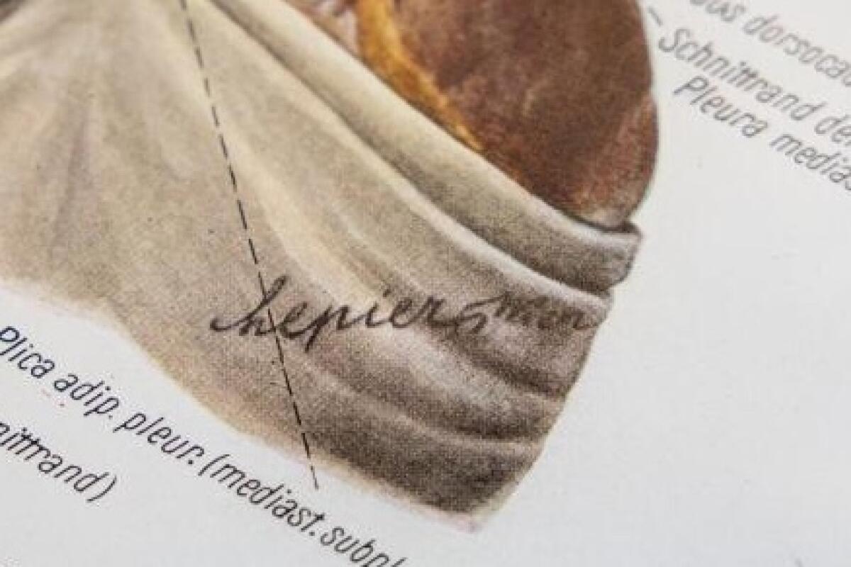
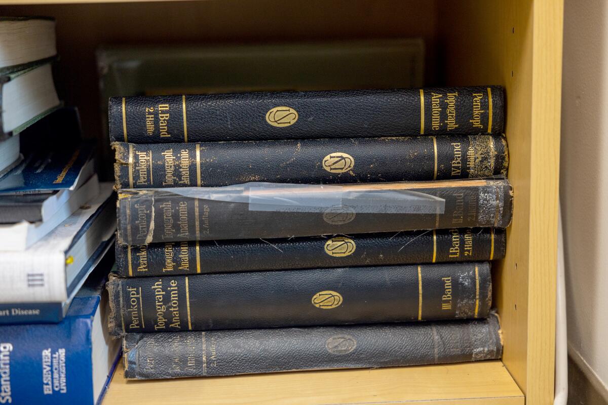
1. A stack of volumes of the Pernkopf atlas on a shelf in Dr. Kalyanam Shivkumar’s UCLA office. 2. Erich Lepier, one of the Pernkopf atlas illustrators, repeatedly included a swastika after the cursive R in his signature. (Allen J. Schaben / Los Angeles Times)
Yet use of the atlas persisted. Hildebrandt said that a decade ago, dental students in her classes “were basically giving each other thumb drives with bootlegged copies of the head and neck.”
Other anatomical atlases exist, but these illustrations had especially fine details, including of the nerves extending beyond the brain and spiral cord. One survey of nerve surgeons found that 13% of respondents were using the atlas. Among those who have publicly grappled with it is Dr. Susan Mackinnon, a surgery professor at Washington University School of Medicine in St. Louis known as a pioneer in nerve regeneration.
“I used this textbook for years before I knew the history of it,” she said. “My brain is contaminated with that. I can’t undo that.”
Mackinnon sought ethical guidance. Rabbi Joseph Polak, a Boston University assistant adjunct professor of health law who survived the concentration camps as a child, said one dilemma involved a patient in excruciating pain.
Polak recalled that the patient had told Mackinnon that “if you can’t find the nerve to stop the pain, then I want my leg amputated.” The rabbi walked through Jewish teachings that applied to the ethical quandary and conferred with other experts, penning a set of recommendations called the Vienna Protocol.
Among his urgings to doctors: If you use these drawings, make it clear to patients where they came from.
The Third Reich wanted “to extinguish them and to extinguish eventually all memory of them,” the rabbi said of Holocaust victims, speaking at a recent forum about the atlas. But when a doctor tells patients about what happened to the people depicted in the drawings, he said, “they’re being called out of that darkness.”
Mackinnon now keeps the atlas locked away. In the rare cases she feels she needs to consult it to operate, she tells patients and co-workers about the man behind it. His firings of Jewish doctors. The grim details in its pages — shorn hair, emaciated bodies — that began to raise suspicions about its terrible origins.
The only reason to use it, she said, is to save someone from misery — and only if “nothing else will help you.”
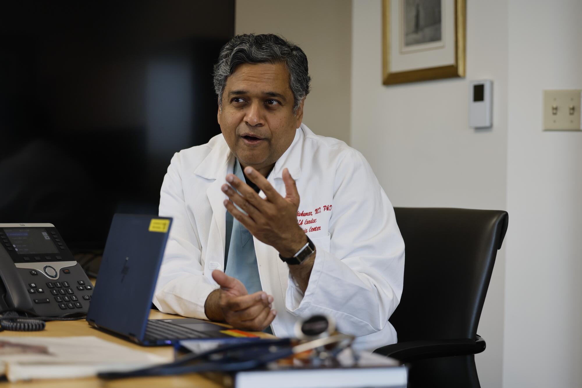
“Could we be better” than Pernkopf? Shivkumar asked. “Could we not be making something that’s completely untainted?”
(Allen J. Schaben / Los Angeles Times)
Shivkumar said his goal is to eliminate the need to consult those pages at all. Inside UCLA’s Center for the Health Sciences in Westwood, he showed off a donated heart, prepped and ready for its close-up in a corner of the lab outfitted with a black backdrop and brilliant lights.
A spent heart normally wilts like a deflated balloon, but this one had been pumped with chemicals to imitate the fullness of life. The team first puts the organs to use in research, then carefully dissects them for imaging.
Bringing out a bisected piece of a heart, Dr. Shumpei Mori displayed how its inner architecture could be captured on camera, threading a catheter through the organ as a co-worker snaked in an endoscope.
“The internal structure is really fine and delicate,” said Mori, a specialist in cardiac anatomy who had jumped at the chance to do something new in the field.
“Even Pernkopf simplified the anatomy” in its drawings, Mori said. “What we are doing is more complicated.”
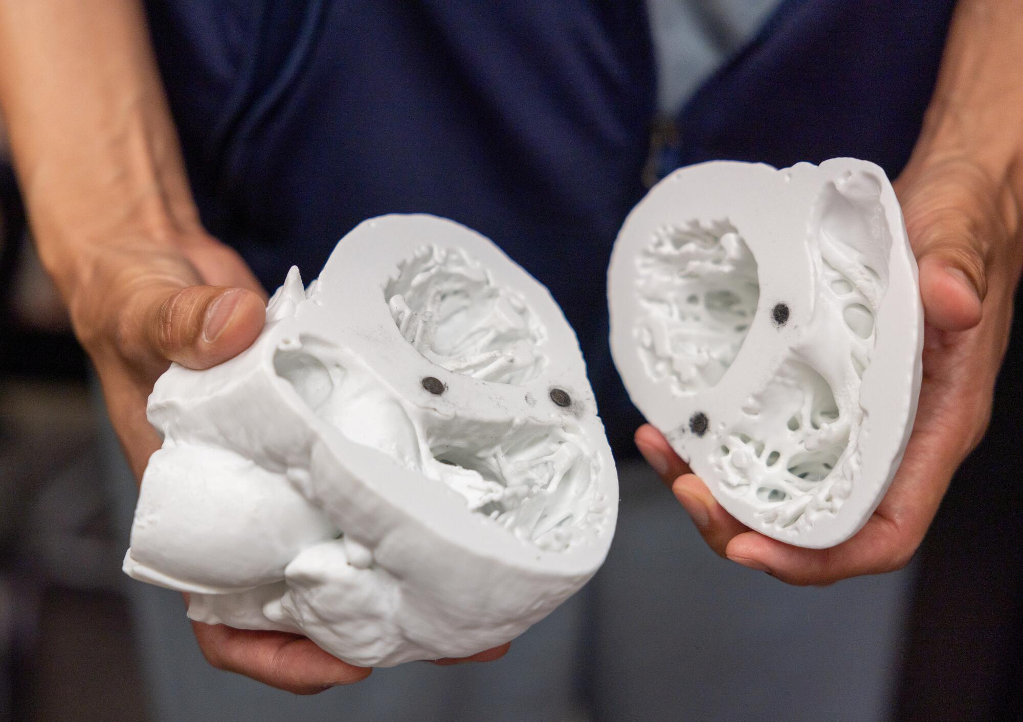
Dr. Shumpei Mori holds a detailed model of a heart. “Even Pernkopf simplified the anatomy,” he said. “What we’re doing is more complicated.”
(Allen J. Schaben / Los Angeles Times)
The camera is far from their only tool: The team has generated 3-D images to illustrate the dimensions of the inner structures of the heart; done CT scans to produce hand-held models; and used sophisticated imaging from a microscope to reveal the lattice of nerves connecting to the organ — part of the signaling system that Shivkumar calls “the internet of the human body.”
In another lab, Mori carefully unzipped a bag on a metal gurney to reveal the stripped-down interior of a cadaver diligently dissected over a year and a half, its rib cage cracked open like a weighty book. Shivkumar pointed out the pale web of nerves stretching up through the neck. Mori had painted them yellow by hand.
The human body might seem like well-traveled territory, but as physicians work to find less invasive ways of healing, such as attacking a cancer with ultrasound, Shivkumar said there is “a volcanic desire for this kind of information.” Snip the right nerve, he said, and you can avert the need for a heart transplant.
“Pernkopf never did nerves like this,” he said with pride.
Amara Yad is also an act of “moral repair” meant to honor the victims, said Dr. Barbara Natterson-Horowitz, a UCLA cardiologist and evolutionary biologist who helped support the project. The Nazi atlases “were like documents of death. The atlases that Shiv is creating are really living, interactive tools to support life.”
When Shivkumar decided to launch the project, he had been inspired by the words of USC emeritus professor of rheumatology Dr. Richard Panush, who had pushed to set the atlas aside in the library of the New Jersey medical center where he had worked, moving it to a display case that explained its history.
Panush said the old atlas should be preserved only as “a symbol of what we should not do, and how we should not behave, and the kind of people that we cannot respect.”
Doctors need to know that history to understand their own moral fallibility, Hildebrandt said. Physicians in Nazi Germany “still thought they were doing the right thing,” she said, even as they failed to see some people as human.
Rabbi Polak stressed that doctors at the time “had the deepest, most profound respect of the masses.”
Yet when the Nazis took power, “it turned out that a vast proportion of them were moral sleazeballs,” Polak said. “They were the first to join when they saw that it could promote their careers.”
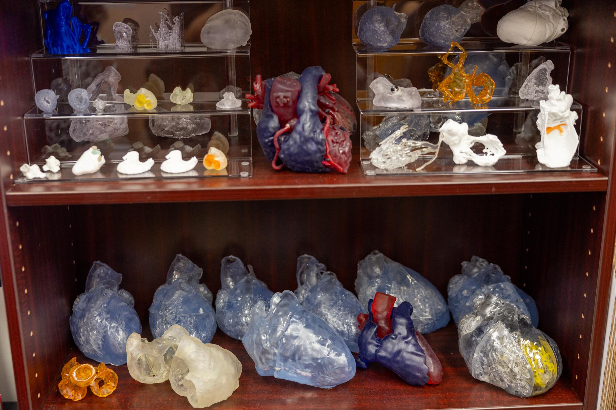
Model hearts line a bookshelf at UCLA. The Amara Yad project is working with other universities to tackle other parts of human anatomy.
(Allen J. Schaben / Los Angeles Times)
Shivkumar said that beyond making new tools for physicians, the Amara Yad project is working with Oxford University to develop an accompanying curriculum that will explore ethical failures in medicine. Pernkopf’s anatomy book is only one example.
The history of the atlas “invites the contemplation of how doctors and medical scientists and anatomists are related to a regime,” said Sari J. Siegel, who heads the Center for Medicine, Holocaust and Genocide Studies at Cedars-Sinai. Thinking about it underscores that “medicine is political.”
“It can’t be divorced from the larger contexts in which it exists.”
Shivkumar, born to a Hindu family in the southernmost state of India, is used to people wondering why he became “possessed” with this project. He recalls first learning about the Holocaust from a photographer friend of his grandfather, a former newspaper editor once imprisoned for sedition against the British Empire.
He was 11 when the photographer showed him images dating to World War II, and it chilled him “to see that human beings could be so brutal to other humans.” As a child, his parents had told him they owed the world because their part of India was lucky to be long spared from such conflict.
In Amara Yad, we “get a rare opportunity in history to correct an unbelievably depressing stain that was placed in our field,” he told the Heart Rhythm Society.
It irritates him to think of the abundant resources that a Nazi had at hand to do this sort of work. “Imagine having five Shumpeis!” he exclaimed at one point, gesturing at his colleague who hand painted the nerves. At UCLA, the project has piggybacked on ongoing research and relied on donations. He is hoping to garner $500,000 annually to continue and expand the work.
But Shivkumar likes to quote the Emperor Ashoka on that point: “To do good is difficult. One who does good first does something hard to do. … Truly, it is easy to do evil.”

Federal employees who were axed during waves of cuts by the Trump administration have fought back against the dismantling of a key climate science website, Climate.gov, and put up a new site, Climate.us, that can now do everything the original did.
The site, with millions of users each year, was known for colorful charts that anyone could freely download and that simplified giant sets of data, such as temperature readings. Now it refers to another page and is no longer being updated.
Daniel Swain, a UC Agriculture & Natural Resources climate scientist, called the resources available at Climate.gov “the most efficacious dollars spent by NOAA on public-facing science, possibly ever.” He has used graphics from the former website on his popular weather blog.
“I am a terrible artist or illustrator. It would be very bad if I had to create those on my own.” Swain said. The website didn’t just make graphics that were beautiful, he said, they were accurate and reliable because of the network of researchers who fact-checked them.
Rebecca Lindsey was the editorial lead and program manager for Climate.gov until February 2025, when her position at the National Oceanic and Atmospheric Administration was eliminated by the Elon Musk-led Department of Government Efficiency, or DOGE. She explained that the online resource was “a bridge between scientists, data and the public.”
Lindsey and her team have now rebuilt the bridge piece by piece, if just a bit further downstream.
The team is made of the same editorial and technical staff that ran Climate.gov. It’s paid for through a crowdfunding campaign and one large, anonymous donation.
The group has raised some $380,000, about $100,000 of which came in the last week. They also have recruited 80 scientists who are willing to volunteer as subject matter experts and fact checkers. It’s enough to keep the work going through February while they seek more long-term funding.
The first iteration of Climate.us went online in 2025 to keep the last 15 years of work from the government website available. The newest version restores the full function of the previous website.
For Californians, the timing could be important.
“We’re headed for a very strong El Niño event that will have significant implications for Southern California,” Swain said. “Climate.gov and the scientists behind it did a great job walking people through the last one, and I would expect that’s the case this time as well.”
Climate.gov excelled at tapping into a pool of academic experts to explain what was happening in nearly real time. This allowed the public to see how events such as wildfire, drought or large weather patterns such as El Niño were shaping their lives when they needed the information most. Research from academic institutions, by contrast, can take years to publish results from major natural disasters.
Swain emphasized that cuts to resources that give context to hard-to-interpret data is not just a loss for the research community.
“It’s getting more and more difficult for the American public to access the science and the scientists that their tax dollars have supported for over half a century,” he said.
With the revival of Climate.us, Swain said he plans to directly use the site and its graphics to keep Californians connected to the world of climate science.

Scientists have long dreamed of discovering the alchemy by which chemicals can be turned into life. On Wednesday, a team at the University of Minnesota announced that it had taken a major step toward that vision.
Blending together dozens of ingredients, the researchers have synthesized simple cells that feed, grow, reproduce and compete with one another for food. If these cells are not yet fully alive, they have most of the hallmarks of life.
“Life is not binary,” said Kate Adamala, a synthetic biologist who led the research. “That’s why I’m hesitant to call this ‘alive.’ There’s no clear line, as much as we would love it to be.”
Until now, scientists had never mastered the recipe for a cell that can perform so many functions, said John Glass, a synthetic biologist at the J. Craig Venter Institute in La Jolla, Calif., who was not involved in the study.
“It is dazzling that she has put these things all together,” he said.
Drew Endy, a synthetic biologist at Stanford University, said, “It’s a cell that was built, not born. It’s constructed, but it does what cells do.”
Dr. Adamala named her creation SpudCell, after its potato-like appearance. Rather than patent it, she and Dr. Endy are organizing a community of scientists to focus on making SpudCells more fully alive and adapting them to new kinds of experiments.
They and their colleagues have founded a nonprofit research organization that Dr. Endy estimates will spend hundreds of millions of dollars on the effort in the next decade. Hundreds of scientists are expected to join.
“We’re going to remember this moment,” said Roseanna Zia, a computational biologist at the University of Missouri who was not involved in the project.
Dr. Adamala and her colleagues posted a 190-page account of their work online. The research is under review for publication in a scientific journal.
Scientists hope synthetic cells can tell them things about life that natural cells cannot, including such basic questions as how many genes are necessary for a minimal form of life.
But synthetic cells also might someday be engineered to do things that natural cells can’t, like making new kinds of medicine or drawing large amounts of carbon dioxide from the atmosphere. In theory, engineered SpudCells might produce a vast range of proteins that natural cells cannot be coaxed to make, or even toxic chemicals like rocket fuel.
Now, “we can think about doing chemistry that we’re barely getting our heads around,” Dr. Glass said.
The trouble with life as we know it: mysterious, messy complexity. Our own DNA contains tens of thousands of genes, as well as millions of molecular switches turning those genes on and off. Scientists barely have a clue as to what many of those pieces of DNA are doing. Often a gene that they think they understand turns out to be performing other jobs than scientists expected.
One way to sidestep this intricacy is to simplify.
In the 1990s, a team led by the late biologist Craig Venter began studying a microbe that had fewer than 1,000 genes. The team, now led by Dr. Glass, went on to strip the microbe’s genome down to 525 essential genes.
In a 2016 paper, the team reported it didn’t know what a third of those genes were doing. Dr. Glass and his colleagues have spent the last decade trying to solve the puzzle, and they still can’t say what 56 of them do.
“There are still significant tasks that every cell has to do that we don’t know,” Dr. Glass said.
Other researchers tackled the problem from the opposite direction. Instead of working from the top down, they moved from the bottom up, seeking to combine lifeless molecules to produce a living cell.
Since the 1990s, several labs have bitten off small pieces of this problem. Some of them have perfected recipes to make hollow bubbles from oily molecules. Others have found ways to encapsulate simple genetic molecules inside those bubbles.
But scientists struggled to put these pieces together into more complex systems, let alone something that could be called a cell.
In recent years, Dr. Adamala took on one of the fundamental challenges: cell division. A natural cell divides with the help of proteins that lock together into a ring anchored to its inner wall. The ring winds itself tighter, pinching the cell in two.
Other proteins act like winches, moving DNA and other molecules into the forming cells, so that they have the ingredients necessary to keep living.
At first, Dr. Adamala tried building a simpler version of the natural system. But then she decided not to mimic real cells at all.
Biophysicists had found that if they stuck proteins on a membrane, they created pressure that made the membrane bend. Dr. Adamala and her team created bubbles that could snag proteins floating around them. When a bubble collected enough proteins, its surface began bending inward until it popped in two.
While the idea was simple, getting it to work in the lab required a year of experiments. “But once it works, it works,” Dr. Adamala said.
That success prompted the team to try to build a synthetic cell in its entirety.
The first step was to create a broth of the molecules necessary for a cell to operate. The recipe ultimately included about a hundred kinds of proteins and simple molecules required for crucial chemical reactions, such as making new proteins from genes.
The researchers also provided their synthetic cell with genes borrowed from a virus and the ubiquitous microbe Escherichia coli. They picked 36 genes for basic jobs like copying DNA.
After mixing these ingredients together into a soup, the scientists added the building blocks of membranes. They spontaneously joined together into bubbles, each engulfing some of the soup.
Many of these bubbles ended up encasing the right mix of genes, proteins and other molecules, and they started carrying out the chemical reactions seen in real cells.
As the new cells floated in flasks, Dr. Adamala and her colleagues added food. The cells slurped up small molecules through channels on their surfaces.
The scientists also put in small bubbles loaded with proteins and other molecules that were too big to fit through the channels. By bumping and fusing into one of these bubbles, the cell could feed on the treats inside.
As the cells fed, they grew. And in just a few hours, they were big enough to divide.
The scientists added a special protein to the flasks, which latched onto the surface of the cells and forced them to bend inward. Once the cells split in two, the pair of new cells kept growing.
Now the SpudCells grew, fed and reproduced. As it turned out, the cells even had a rudimentary ability to evolve.
Dr. Adamala and her colleagues created a mutant version that bound more tightly to the snack-filled bubbles floating around it. To test it, they created a 50-50 mixture of original and mutant SpudCells.
The cells competed for five generations for food. Eventually the mutants outnumbered the originals, suggesting that they were outcompeting the originals for food.
“That’s the shake-the-ground accomplishment here,” said Dr. Zia. Scientists will be able to put various synthetic cells in competition with one another and rapidly develop more sophisticated ones.
For all this evidence of life, SpudCell still has some major shortcomings. For starters, it can’t make the molecular factory that produces new proteins, called a ribosome. The cells can carry all the genes they need to build ribosomes, but for some reason the parts don’t come together.
For now, Dr. Adamala and her colleagues have to feed ready-made ribosomes to SpudCells. This solution has an expiration date, though: SpudCells can keep making proteins through five to 10 generations before they fail as their ribosomes become defective.
“I don’t want to say it dies, but it stops working,” Dr. Adamala said.
When Dr. Adamala showed SpudCell to Dr. Endy last year, he was so awestruck that he decided to help her found Biotic, the nonprofit organization intended to create a community of SpudCell researchers.
“I’m pouring my life’s work into this,” Dr. Endy said. One of the first tasks for Biotic will be to make it easier for other scientists to create SpudCells.
Dr. Adamala can create a fresh batch of them in her own lab in about a day. But that’s only because she has freezers full of purified proteins and an intimate understanding of each step of her recipe. Biotic expects to offer scientists easier recipes and provide the required ingredients.
Dr. Endy hopes that the open-source tools will encourage scientists to collaborate on building new kinds of SpudCells with more of the defining features of life, such as the ability to make their own ribosomes and to divide indefinitely.
“It’s completely doable,” said Dr. Glass.
Biotic researchers are already planning their first meeting, in September in Philadelphia. High on their list of priorities will be formalizing plans to safeguard this area of research.
For now, the synthetic cell can only survive a few generations on a special lab diet. But future versions may be more robust, raising the possibility that someone might someday use SpudCells unethically, perhaps even to make a weapon.
Dr. Endy argues that an open-source research community will be better prepared to prevent that from happening. “We can have these conversations now, as opposed to waiting for somebody else to do it, and then we’re just all reacting,” he said.
Dr. Endy likens SpudCells to a biological version of the Wright flyer, the crude plane that the Wright Brothers used to make the first sustained controlled flight in 1903, ushering in the age of airplanes.
“The Wright flyer flying for 12 seconds doesn’t get you a 737,” Dr. Endy said. “This is just the beginning.”

For the next five years, the Environmental Protection Agency has indicated it will not require public water utilities to test for microplastics or pharmaceuticals in drinking water, according to a proposed rule published in the Federal Register.
On Friday, the EPA submitted a list of chemicals it plans to test for under the Unregulated Contaminant Monitoring Rule, a mandatory testing program used to collect information about concerning chemicals in drinking water that could be harming human health. It did not include microplastics or pharmaceuticals.
The omissions come after announcements by EPA Administrator Lee Zeldin earlier this year that his agency was designating microplastics and pharmaceuticals priority contaminants for testing.
“This is a direct response to the concern of millions of Americans who have long demanded answers about what they and their families are drinking every day,” he said at an April news conference with Health and Human Secretary Robert F. Kennedy Jr. at EPA headquarters.
Zeldin’s announcement was seen at the time as a move to placate the increasingly disgruntled Make America Healthy Again contingent of Trump supporters.
Now the agency says it has no validated or standardized method to test for the plastic particles in drinking water, and wouldn’t be able to develop one before December, when testing is required to begin.
Among the 33 chemicals the EPA will require water utilities to test for are seven PFAS, or forever chemicals, and three pesticide residues.
It will be five years before the EPA proposes another list.
The EPA did not respond to a request for comment.
The agency noted in its proposed rule that it will collaborate with other federal agencies to “evaluate risks and exposures” of microplastics for future monitoring.
Environmentalists reacted with frustration and resignation. They pointed out that the European Union has developed methods to test for the tiny plastic particles, which have been found in people’s blood, brains and lung tissue. California has one in the works.
“The California water board has spent a lot of time and money on how to measure in drinking water,” said Judith Enck, a former EPA regional administrator and president of the anti-plastic environmental group Beyond Plastics. “EPA should give them a call.”
California was required by a 2018 state law to establish a protocol for local water utilities to test for the particles in drinking water. The state has not yet begun reporting its results, but protocols were established in 2021. Blair Robertson, a spokesman for the State Water Resources Control Board, said it’s not “a fully validated, end-to-end regulatory method” yet.
At the April meeting, Zeldin announced that he would place microplastics on what is known as the Contaminant Candidate List, which acts as a preliminary “watch list” of unregulated, priority contaminants in drinking water. Like the mandatory monitoring list, it is updated only every five years. The most recent list was published on April 2 — the day he made his announcement.
“Americans have been ignored as they sound the alarm about plastics in their drinking water,” Zeldin said during the announcement. “That ends today by placing microplastics on the contaminant candidate list for the first time ever. EPA will follow the science, will pursue answers and will hold ourselves to the highest standards to protect the health of Americans.”
There appears to be no clear association between these two lists, although the contaminant list is supposed to inform the monitoring list. Seventy-five chemicals and four chemical groups (microplastics, pharmaceuticals, PFAS chemicals, and disinfection byproducts) were listed on the 2026 contaminant list. Only seven of those chemicals were also on the proposed monitoring list (as well as seven PFAS chemicals).
When Zeldin announced microplastics as “‘a priority contaminant for regulation,’ and called it ‘a historic action on microplastics,’ he made it seem like the administration was going to take microplastics seriously,” said Mary Grant, water policy director for the environmental group Food & Water Watch.
“By not including them, they made it clear they don’t actually have plans to immediately address this crisis by getting the real-world monitoring data that we need right now to really start correcting ourselves,” she said.
Craig Davis, senior director of plastics chemistry at the American Chemistry Council — the nation’s largest trade group for chemical companies — said that while his organization supports microplastic research, it also agrees with the EPA’s decision not to include them in the monitoring list.
“National drinking water monitoring should be based on validated, standardized methods that can produce reliable and comparable data,” said Davis in a statement. He said “limited” national monitoring resources should be focused where data can produce “actionable public health information.”
The public has 60 days to comment once the plan is published in the Federal Register.
-

 Health5 minutes ago
What Is Retatrutide? Dr. Dubrow Calls It the Most Powerful Weight-Loss Drug
-
 Lifestyle20 minutes ago
How World Cup fans reflect America back at us : It’s Been a Minute
-

 Technology28 minutes ago
Apple’s entry-level MacBook Pro could be up for a redesign
-

 World35 minutes ago
Khamenei body in cold storage as feared Basij mobilizes ahead of historic Iran funeral
-

 Politics38 minutes ago
Coalition of 25 states sues Trump admin over Medicaid work rule designed to prevent fraud
-

 Health43 minutes ago
West Nile virus detected in southern state as health officials warn residents about mosquitoes
-

 Sports50 minutes ago
USA World Cup star Folarin Balogun receives controversial red card during Round of 32 match
-

 Technology53 minutes ago
A missing kitten rode under a car hood. AI brought her home